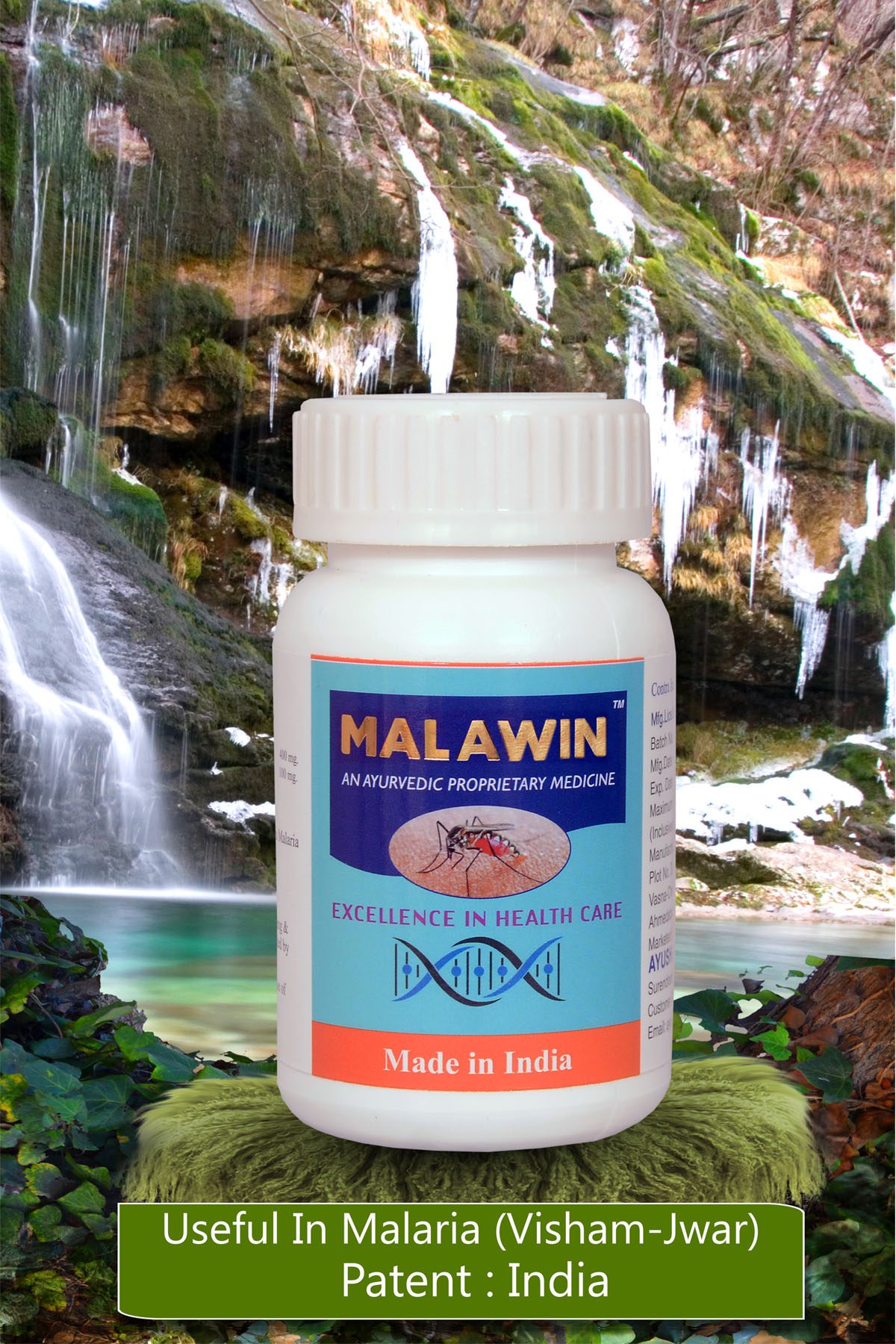
Description
CLINICAL INDICATIONS: Helps to control Malarial Conditions.
OBJECTIVE OF NOVEL DRUG COMPOSITION
Forty one percent of the world’s population live in areas where malaria is transmitted (e.g., parts of Africa, Asia, the Middle East, Central and South America, Hispaniola, and Oceania). In areas of Africa with high malaria transmission, an estimated 990,000 people died of malaria in 1995 – over 2700 deaths per day, or 2 deaths per minute. Over 3 billion people live under the threat of malaria. It kills over a million each year. An estimated 350-500 million people suffer from malaria every year. Malaria occurs in over 100 countries worldwide Malaria caused 10.7% of all children’s deaths in developing countries. Malaria is caused by four species of parasitic protozoa that infect human red blood cells. Protozoa are one-celled organisms that are as sophisticated as a human cell. Malaria parasites feed on red blood cells for a living. Malaria parasites have a complex life cycle. In order to live, they need to have both a human and a mosquito host.
Indian Scenario:
Of the 5 million confirmed cases of malaria which are reported each year from countries outside of Africa, nearly 3 million are from India and Pakistan. India reported 1.86 million confirmed cases and 1000 deaths in 2003. 45 percent of these cases, or about 850,000, were plasmodium falciparum.
In India the risk of contracting malaria is unevenly distributed across the country; 20 percent of the population is reporting 80 percent of the cases. 80 percent of the India’s population now lives in areas with low incidence of malaria. Orissa has the highest number of cases and deaths in the country. Other high disease burden states include Gujarat, West Bengal, Chhattisgarh, Madhya Pradesh, Rajasthan, Uttar Pradesh, Karnataka, Jharkhand, and Maharashtra. The Northeast states not only have a high burden of malaria, but also a significant share of Plasmodium falciparum and drug resistance.
The Global Warfare against the malarial parasite:
Forty years ago, the nations of the world embarked on a worldwide effort to eradicate the parasite that causes the disease malaria. The effort ended in failure 17 years later. At the beginning there was a belief that given enough insecticide, medicine and money, the parasite could be eliminated from the planet. It gradually became apparent that the strength of the parasite to survive and continue causing disease in human hosts had been grossly miscalculated. Analyzing the strategy and results of the eradication effort has been useful in improving public health policy of malaria control. The lessons learned are important because they apply to science and politics as well.
CONVENTIONAL TREATMENT IN MALARIA:
Many conventional drugs e.g. Quinine preparations are available at present. But most of them have side effects or adverse effects like, blurred vision, loss of appetite, liver dysfunction, deafness, drowsiness, vomiting, rolling, ulceration, gastrointestinal discomfort or pain, nausea, floating scotomata, mild cyanosis and Retinopathies.
REF.: 1. World Health Organization 1966
2. Douglas R. G.: Proceedings of a symposium: Anti-Virals, 1980-82
AM.J.Med.
3. Vagbhatt : Astang – Hridaya
4. Shukla M. H. : Clinical summary of anti-Malaria Drug
(Bioactive Formulations) 1992-2012
LIFE CYCLE IN MAN: Exoerythrocytic Schizogony:
The next stage of life cycle, the primary exoerythrotic schizogony, begins in the tissues of the host. This pre-erythrocytic (PE) phase develops in the parenchymatous cells of the liver. On entering the cell, the sporozotie rounds up and the necleus undergoes repeated division. In the course of 6-16 days the schizonts matures and divides to produce small single-nucleated “merozoites”. The liver cells ruptures and the merozoites escape either into the blood stream where they invade erythrocytes and initiate the erythrocyte (E) phase, or into contiguous liver cells where they may initiate a secondary exoerythrocytic phase (EE).
In falciparum infection on secondary infection of the liver cells occurs, but in the other infections this takes place and the liver phase (EE) persists. This eventually provides the mechanism for relapses. Only a single generation of primary exoerythrocytic schizogony is required before the merozoites entering the blood stream are capable of invading the erythrocytes and initiating the sexual blood cycle of the parasites, the E phase.
It should be informed hereby that the pattern of parasites in patients with ovale malaria is:
Falciparum malaria: E-phase only.
Other malaria: E-phase plus persistent, EE phase.
In P. falciparum infection, renewed manifestations of the malaria subsequent to the primary attack, if not resulting from fresh infection, are due to multiplication in the blood of existing E phase parasites. This is a recrudescence, not a relapse. Relapse is explained on the ground that merozoites are discharged at irregular intervals into the blood stream. As the blood infection progresses, immunity becomes established and the merozoites are destroyed. The immunity, however, has no effect on the tissue parasites. GAMOCYTES are pathologically inert. It is the E phase alone which is responsible for the clinical and pathological disturbances in the host which constitute malaria. Functional damage to vascular endothelium leads to escape of protein and water and than increase in blood viscosity and finally stasis.
Changes in the brain are common in falciparum malaria. At autopsy in so called “cerebral” malaria; the capillaries are seen to be loaded with erythrocytes, many infected with schizonts and lying along the endothelial membrane.
Renal lesions develop in acute falciparum infections and black water fever. The bone marrow in acute falciparum malaria is hyperplasic, representing compensatory normoblastic activity but there is usually no increase in reticulocytes in the peripheral blood until some days after treatment has removed the parasites. In very severe infections there may be no erythropoietic response and the marrow is hypoplastic. Myeloblastic activity is usually depressed, as indicated by a granulocytic leucopenia.
The unconcentrated urine has a low specific gravity, which may be masked by the presence of protein or hemoglobin. The plasma protein is unaltered or may be lowered in the early stages of an acute infection. As the disease proceeds, the protein content may increase as a result of increase in gamma globulin despite a fall in albumin resulting from liver dysfunction and the albumin/globulin ratio is correspondingly reversed. In the late stages of severe falciparum malaria the blood sugar may be very low.
Respiration is rapid. The patient complains of throbbing headache. He is restless, excited and euphoric, he may become disoriented or pass into delirium.
The commonest forms of cerebral malaria are (In complicated falciparum/pernicious malaria), hyperpyrexia, gastrointestinal complications and algid malaria. Black water fever is regarded as a complication of falciparum malaria. Other features of malaria attack, including anemia, splenic and hepatic enlargement are present and progressive. Diarrhea is common and the comatose patient is usually incontinent.
Malaria, especially P.falciparum infection may cause abortion at any stage and premature labour in the last trimester especially in primapara. In the late stages of pregnancy and following childbirth, mothers in holoendemic or hyperendemic areas of falciparum malaria lose some of their acquired immunity and may suffer from severe attacks which are sometimes fatal if not treated timely. In highly endemic areas P.falciparum infected women often come to term with severe anaemia, which may be derived from mixed iron deficiency and folic acid deficiency. In some cases, an enlarged spleen or liver may obstruct the expansion of the uterus as the fetus develops and lead to abortion or difficult delivery.


